Previous Issues Volume 9, Issue 10 - 2024
Oral Rehabilitation Case enhanced by Plastic Periodontal Surgery using Sub-epithelial Connective Tissue Grafting
Ola Khairallah1, Aseel Al-Mashout1, Aous Dannan2,*
1Post-graduate student, Faculty of Dentistry, The International University for Science and Technology, Ghabagheb, Syrian Arab Republic
2Professor and Head Department of Periodontology, Faculty of Dentistry, The International University for Science and Technology, Ghabagheb, Syrian Arab Republic
*Corresponding Author: Prof. Aous Dannan, Professor and Head Department of Periodontology, Faculty of Dentistry, The International University for Science and Technology, Ghabagheb, Syrian Arab Republic, Phone: 00963 933300 485, Email: [email protected]
Received Date: October 05, 2024
Published Date: November 13, 2024
Citation: Khairallah O, et al. (2024). Oral Rehabilitation Case enhanced by Plastic Periodontal Surgery using Sub-epithelial Connective Tissue Grafting. Mathews J Case Rep. 9(10):192.
Copyrights: Khairallah O, et al. (2024).
ABSTRACT
Comprehensive oral rehabilitation cases are considered to be a major challenge for dentists, as they involve the use of various specialties to achieve the optimal functional and aesthetic results that the patient aspires to. Various periodontal surgeries play an important role in adding an indispensable aesthetic touch to comprehensive rehabilitation cases, especially when combined with the construction of well-crafted cosmetic prostheses. This clinical case presents a comprehensive oral rehabilitation process for a 54-year-old male patient, and the various steps that were carried out through various dental treatments were explained. The focus was also on the process of rebuilding the missing interdental gingival papillae from the anterior region as a major part of that case, with an explanation of its significant impact on adding a high aesthetic spirit in the end.
INTRODUCTION
Achieving optimal results in comprehensive oral rehabilitation usually requires collaboration between different dental specialties. Periodontal treatment is usually the first step in such cases, followed by conservative and endodontic treatments and extraction of hopeless teeth or irreversible decayed roots, and these cases end with giving the patient a new occlusal function through crowns, bridges, dental implants or removable appliances. Some periodontal surgical procedures play an important role in restoring the lost aesthetic aspect in these patients, such as surgical crown lengthening, cosmetic gingival resection and trimming, in addition to gingival tissue grafting procedures to cover gingival recessions or reconstruct missing interdental gingival papillae, which pose a major challenge, especially in the anterior region [1,2].
The presence of interproximal spaces may result in aesthetic and phonetic problems. Interdental papillae can be lost as a result of several distinct clinical situations [3]. The most common reason in the adult population is loss of periodontal support because of plaque-associated lesions and previous pocket elimination periodontal therapy. However, abnormal tooth shape, improper contours of prosthetic restorations, and traumatic oral hygiene procedures also may produce negative changes on the size and shape of the interdental soft tissues [4]. Kandaswamy et al. reported that dark triangles (or black holes) are more likely to develop following labial movement of overlapping or palatally placed incisors and diastema closure [5].
Several surgical and nonsurgical techniques have been proposed to treat soft-tissue deformities to manage tissue changes in the interproximal space. The nonsurgical approaches include such adjunctive therapies as orthodontics and both prosthetic and restorative procedures to modify the interproximal space, thereby producing subsequent modifications to the interdental soft tissue [3,4,6].
Surgical techniques aiming at correcting the “black hole problem” have relied mainly on the use of sub-epithelial connective tissue grafts [4], repeated interproximal curettage [7], or displacement of the interproximal palatal tissue in the buccal direction [8]. However, specialized periodontal surgeries in the aesthetic fields cannot give the desired results alone, but rather require technical support in the field of cosmetic reconstruction of the teeth (i.e., the pattern and model of crowns or bridges and their relationship to the gingiva).
The following clinical case presents a series of dental procedures aimed at rehabilitating complete oral function in a patient in his fifties, with emphasis on a specialized periodontal surgical procedure that reconstructed the missing interdental papillae in the anterior region.
CASE DESCRIPTION
A 54-year-old male patient visited the College of Dentistry clinics at the International University for Science and Technology (IUST) complaining that he could not chew food well, as well as some aesthetic concerns. The patient had a controlled high arterial pressure (hypertension). After clinical examination, it was found that there was severe abrasion of the anterior teeth due to muscle trismus and excessive destruction of the posterior teeth which led to loss of masticatory function. Old bridges, gingival recession, periodontitis, and exposure of some roots were also notable (Figure 1).
Figure 1. Intra-oral pre-operative situation of the case.
After radiological examination, it was found that there was a considerable bone resorption in the entire oral cavity (Figure 2).
Figure 2. Panoramic radiography before treatment, in which bone destruction, revealing periodontitis, is evident.
The upper teeth, especially the anterior area, showed absence of interdental gingival papillae, coinciding with the absence of contact points between teeth. This led to an unesthetic appearance (Figure 3).
Figure 3. Situation after scaling and root planing. Absence of papillae between anterior teeth is evident.
An appropriate treatment plan was set to be implemented during a specified period of time under the supervision of an elite group of doctors.
Firstly, in order to control the periodontal status, scaling and root planing were carried out to reduce bacterial virulence by using an ultrasonic scaler device and Chlorhexidine rinsing agent at a concentration of 0.12%, finishing with polishing of all surfaces at slow rotating speeds with pumice material. Proper oral hygiene instructions were given to the patient including home teeth brushing, the use of interdental brushes, and flossing. Two weeks later, the periodontal status was re-evaluated and considered as stable and healthy.
In the second step, surgical extraction of teeth 25, 35, and 36 was achieved. Two weeks later, after proper examination, the healing condition was confirmed. Then, endodontic retreatment was started for teeth 17,26,28,37,46,47. Through the treatment and radiological examination of teeth 28,46, it was found that the treatment was not possible due to the presence of lesions and excessive movement, so they were extracted.
On tooth 26, it was noted that there was a lesion on the mesial root with severe resorption in the bifurcation area, with the tooth not showing any mobility, so it was treated through a process of bicuspidation, separating the mesial root and removed it, with endodontic retreatment of the distal and palatal roots (Figure 4).
Figure 4. Tooth #26. (A), Bone resorption in bifurcation area about 6mm. (B), After removal of mesial root and endodontic re-treatment of palatal and distal roots.
After completing the endodontic retreatment, post-and-core restorations were designed for each of the remaining posterior teeth and cemented by zinc phosphate cement.
Old amalgam fillings on teeth 11, 12, 13, 24, 34, 44 were replaced with composite ones, and thus, the anatomical shapes of the teeth were restored.
In the next step, a new smile was designed with appropriate contact points and waxing on the primary cast (Figure 5).
Figure 5. Wax up on primary cast.
Silicone key was taken to produce provisional restoration with Bis-acrylic temporary materials, leaving gaps in the places of the lost interdental papillae to fill them later through a periodontal surgery and to be used as a surgical guide that would guide the healing process later after (Figure 6).
Figure 6. Mock-up provisional restoration made by Bis-acrylic temporary material with inter-dental gaps left.
Up to that point, the patient was ready for a periodontal surgery that encountered the augmentation of lost interdental papillae between front teeth by means of connective tissue graft (CTG) extracted from the hard palate. The whole surgery was performed with the acrylic stent in place. At the first stage, the patient was prepared with verbal support and the necessary preventive measurements were taken, and then deep anaesthesia was performed using Lidocaine/ Epinephrine-free carpule. The greater palatine nerve was anesthetized, and the entire upper anterior area from the right canine to the left canine was also anesthetized by targeting the anterior superior alveolar nerve on both sides. In the next step, the process of extracting the CTG from the palate was initiated by means of a superficial incision made in the dome of the palate on the left side along with the mesial aspect of the first premolar up to the distal aspect of the first molar. Using a mini-scalpel, internal incisions underneath the epithelial layer inside the previously mentioned incision were made and thus dissected the CTG avoiding damage to adjacent tissues and removed it without a full dissection of the epithelial layer. The dimensions of the CTG were about 15*5*2 mm (Figure 7).
Figure 7. The connective tissue graft obtained from the hard palate.
The graft was placed in physiological serum. Then, the bleeding in the dome of the palate was controlled with a continuous suture.
At that time, the CTG was handled by cleaning it from the remnants of adipose or fatty tissues attached to it and then the whole mass was cut into small pieces appropriate to the size of the lost interdental papillae that allowed accepted revascularization (Figure 8).
Figure 8. The connective tissue graft being cleaned and cut it into small pieces.
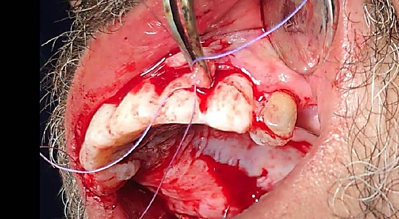
During this period, the CTG small pieces were continuously irrigated with a physiological serum to avoid dying until they were placed in the appropriate area. A suitable vestibular partial thickness flap in the upper anterior region through an intrasulcular incision was raised across the papillae line from the right canine to the left canine without any releasing (i.e., vertical) incisions (Figure 9).
Figure 9. Vestibular flap reflection in upper anterior teeth through intrasulcular incision.
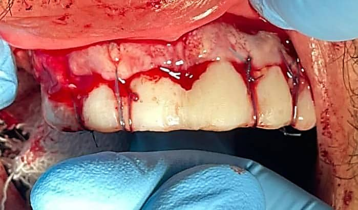
The prepared CTG pieces were placed under the flap on every single place of a lost papilla, and sutured one by one with a (5-0) resorbable suture. After ensuring that the grafts remained in place, the flap was sutured with vertical matrix sutures at each papilla site by using non-resorbable (4-0) nylon sutures (Figures 10 and 11).
Figure 10. Placement and suturing the CTG in papillae regions.
Figure 11. Final suturing after grafting.
Post-surgical instructions were given to the patient such as eating soft foods, avoiding hot food and drinks, and not brushing violently. He was also provided with a medical prescription of oral rinses containing 0.12% chlorhexidine (Biofresh-K®), an antibiotic (Augmentin® 1000 mg.), and a pain reliever (Diclofenac potassium/ Flam-K®) for 10 days.
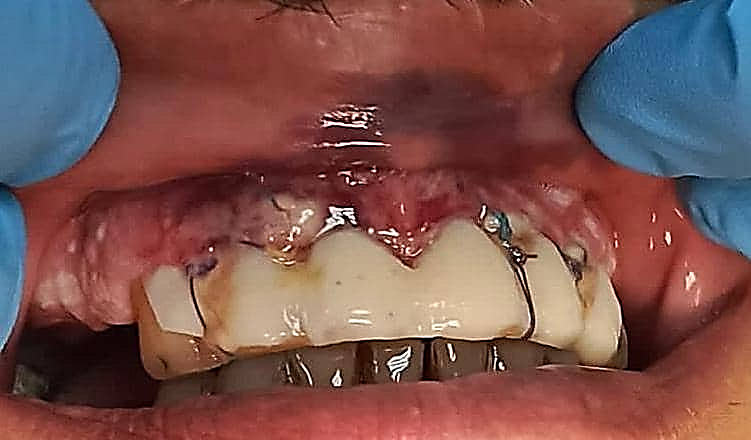
A follow-up of the case was conducted after a week, with an examination of the sutures and grafts to check their stability (Figure 12).
Figure 12. Follow-up after one week.
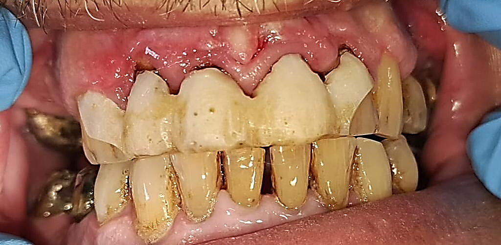
In the second appointment, two weeks later, the sutures were removed and a primary re-building of new interdental papilla was notable (Figure 13).
Figure 13. Follow-up after two weeks with suture removal.
Four weeks later, the provisional restoration was removed and teeth 17, 24, 26, 34, 37, 44, 45, 47 were prepared to receive porcelain-on-metal crowns and bridges, while teeth 11,12,13,21,22,23,31,32,33,41,42,43 were prepared to receive zirconia crowns.
Final impressions were taken with metal trays by means of soft and hard rubber materials. Temporary crowns were placed on all of the teeth prepared at that time. At a later date, the metal crowns were tried on the posterior teeth, and bite wax was taken. At another date, glass ionomer was used as a cement to fix the final restorations after examining the occlusion, fit, angles, aesthetic aspects, and function, as well as the adaptation of new papillae to the new prosthetics.
In the final stages, a removable partial denture (RPD) made of flexible acrylic material was fabricated for the upper right posterior region (Kenedy/ Class III). This type of dentures was chosen due to the presence of bone abnormalities in the form of small protrusions on the top of the alveolar process that could have negatively affect the stability of any removable denture.
Upon completion of the treatment a Night Guard appliance on the lower jaw was fabricated, and the patient was asked to place it in mouth at night for a primary period of 3 months to treat muscular trismus and to protect the prostheses from damage or fracture. However, this stage is not documented in this case report and needs to be demonstrated later.
Final results of the entire case are shown in Figure (14), and the patient was very satisfied with restoring the aesthetic and functional aspects of his intra-oral situation.
Figure 14. Intra-oral photography after cementation of the final crowns and bridges, and design the flexible RPD appliance.
DISCUSSION
Comprehensive oral rehabilitation cases, both aesthetically and functionally, are among the most challenging cases for dentists. Such cases usually include various traditional dental treatments such as conservative treatments, endodontic and periodontal treatments, and then occlusion rehabilitation through crowns, bridges, various restorations, dental implants or partial dentures [9].
Periodontal cosmetic surgeries play an important role in the success of most oral rehabilitation cases due to their positive effect in giving a harmonious and homogeneous appearance to the smile in addition to the blending between the gums and the teeth (white-pink harmony) [10].
The current clinical case presented a comprehensive oral rehabilitation process through sequential and coordinated stages that began with improving the patient's oral health and ended with the completion of fixed and removable prostheses in different areas of the oral cavity. Here we would like to focus a little more on the role of periodontal surgery in the completion of this clinical case. Because of the progressive loss of interdental papillae in the anterior region of the maxilla, a unique technique was used to reconstruct the missing gingival papillae, which was done by removing a connective tissue graft from the palatal vault and then cutting it into small pieces, each of which would later represent a new papilla. After the partial thickness flap was lifted, the connective tissue pieces were placed in their new locations and the flap was then coronally repositioned to its original position.
Reduction or total loss of the interdental papilla may create aesthetic impairments, create phonetic problems, and allow unwarranted food impaction. One of the most difficult and elusive goals for the periodontist in the aesthetic aspect of oral rehabilitation is the reconstruction of the interdental papilla. Periodontal plastic surgery enables enhanced aesthetics in the anterior region where minor surgical procedures can improve gingival contours. One of the goals of periodontal plastic surgery is reconstruction of the lost interdental papilla [2]. However, this is one of the most challenging and least predictable of treatments.
Most previous studies and case presentations contain little or no data regarding short- and long-term results with such techniques [4]. Also, case reports presented in the literature report different clinical situations or reasons for papilla loss. This too may account for the variations of reported success (surgical correction) among techniques as well as reports using the same surgical technique.
Tarnow et al. reported that the distance from the bone crest to the contact point was positively related to the presence of an interdental papilla [11]. When this distance was 5 mm or less, the entire papilla was always present. They found that as the distance increased to 6 mm, the papilla was present 56 percent of the time. When the distance was 7 mm or greater, a papilla was present only 27 percent of the time or less.
In the present case, the mean gain in papilla height was visually accepted and the mean width of the keratinized gingiva increased with no facial gingival recession on adjacent teeth. Comparable findings have been reported in previous case report studies. Carnio [12] reported complete papillary reconstruction using an interposed sub-epithelial connective tissue graft in a case involving a 20-year-old woman with class IV gingival recession and a class III papillary loss between maxillary left central and lateral incisors. He performed a total of three surgical procedures, with identical protocols, in the same area at eight-week intervals.
In another case report, Azzi et al. reported achieving predictable root coverage and papilla reconstruction of class IV recession associated with a loss of papilla using a connective tissue graft [13].
Nemcovsky [14] evaluated the use of a coronally advanced papillary flap in combination with a gingival graft to augment soft tissue in the interdental area in a total of 10 patients. He reported achieving an increase in papilla height in eight patients with no change in two patients due to root proximity and recurrence of a pyogenic granuloma. Post-operative necrosis occurred where the interdental space was very narrow.
In the present case, one positive aspect of having a large interproximal area is the improved blood supply from the flap to the graft. Both the maximized blood supply and the maintenance of papillary integrity by the flap design are essential to avoid flap necrosis and to enhance the grafted tissue. It is important to note that harvesting of the graft was performed just before the surgical detachment of the papilla to prevent the development of a blood clot between the bone and grafted tissue. Blood clots, even small ones, might compromise immediate blood supply to the graft and therefore induce partial necrosis of the transplanted tissue [12].
The success of the procedure used in the current case could be due to the fact that the sub-epithelial connective tissue graft was supported by the coronally advanced flap and the space between the bone and flap was completely filled with connective tissue, which is perfectly stabilized with the aid of simple and tension-free suturing. Since the graft receives nourishment from all directions, the flow of plasma and the in-growth of capillaries from surrounding tissues can be achieved.
Before any periodontal plastic surgery is performed, the dental professional should select the most appropriate technique for each defect to ensure that the patient’s individual needs and complaints are addressed and to achieve the best aesthetic and functional results. The selection of one procedure rather than another depends on a variety of factors, such as the size of the defect (length and width), the width of the keratinized tissue (to allow flap advancement), the amount of connective tissue available from the donor site, patient exposure to risk factors known to influence host response (e.g., smoking), mucogingival phenotypes, and operator experience [15-18].
CONCLUSION
This clinical case presented a comprehensive rehabilitation of masticatory function through a series of conventional dental treatments. The aesthetic aspects, especially in the anterior region, were restored through specialized periodontal surgery that included reconstruction of the missing interdental papillae using a connective tissue graft extracted from the palatal vault with a coronally displaced flap. To the best of our knowledge, there are very few, if any, similar clinical cases in the literature where the technique of dissecting connective tissue grafts into small fragments was used to reconstruct missing interdental papillae, which makes the present clinical case unique. However, the important role of the high-precision manufacturing of ceramic crowns in the anterior region in restoring the aesthetic aspects in an ideal way cannot be overlooked, through paying attention to the details of the locations of the contact points between the teeth and their known effect in returning the interdental papillae to their new positions, which achieves a distinctive aesthetic appearance.
REFERENCES
- McGuire MK. (1998). Periodontal plastic surgery. Dent Clin North Am. 42(3):411-465.
- Miller PD Jr, Allen EP. (2000). The development of periodontal plastic surgery. Periodontol 2000. 11:7-17.
- Kokich VG. (1996). Esthetics: the orthodontic-periodontic restorative connection. Semin Orthod. 2(1):21-30.
- Blatz MB, Hürzeler MB, Strub JR. (1999). Reconstruction of the lost interproximal papilla—presentation of surgical and nonsurgical approaches. Int J Periodontics Restorative Dent. 19(4):395-406.
- Kandasamy S, Goonewardene M, Tennant M. (2007). Changes in interdental papillae heights following alignment of anterior teeth. Aust Orthod J. 23(1):16-23.
- Han TJ, Takei HH. (1996). Progress in gingival papilla reconstruction. Periodontol 2000. 11:65-68.
- Shapiro A. (1985). Regeneration of interdental papillae using periodic curettage. Int J Periodontics Restorative Dent. 5(5):27-33.
- Beagle JR. (1992). Surgical reconstruction of the interdental papilla: case report. Int J Periodontics Restorative Dent. 12(2):145-151.
- Kanany J, Dannan A. (2022). A Comprehensive Aesthetic and Functional Oral Rehabilitation Mediated by Periodontal and Mucogingival Surgeries With A 4-Year Follow Up: A Case Report. World J of Pharma Med Res. 8(5):09-17.
- Al-Halabi R, Al-Hroob K, Dannan A, Al-Nahlawi T, Abd Al-Aal H. (2015). Indirect Composite Laminate Veneers for Upper Anterior Teeth Diastema Closure: A Case Report. Int J Dent Oral Health. 1(4):1-4.
- Tarnow DP, Magner AW, Fletcher P. (1992). The effect of the distance from the contact point to the crest of bone on the presence or absence of the interproximal dental papilla. J Periodontol. 63(12):995-1004.
- Carnio J. (2004). Surgical reconstruction of interdental papilla using an interposed subepithelial connective tissue graft: a case report. Int J Periodontics Restorative Dent. 24(1):31-37.
- Azzi R, Etienne D, Sauvan JL, Miller PD. (1999). Root coverage and papilla reconstruction in Class IV recession: a case report. Int J Periodontics Restorative Dent. 19(5):449-455.
- Nemcovsky CE. (2001). Interproximal papilla augmentation procedure: a novel surgical approach and clinical evaluation of 10 consecutive procedures. Int J Periodontics Restorative Dent. 21(6):553-559.
- Caffesse RG, De LaRosa M, Garza M, Munne-Travers A, Mondragon JC, Weltman R. (2000). Citric acid demineralization and subepithelial connective tissue grafts. J Periodontol. 71(4):568-572.
- Chambrone LA, Chambrone L. (2006). Subepithelial connective tissue grafts in the treatment of multiple recession-type defects. J Periodontol. 77(5):909-916.
- Chambrone L, Lima LA, Pustiglioni FE, Chambrone LA. (2009). Systematic review of periodontal plastic surgery in the treatment of multiple recession-type defects. J Can Dent Assoc. 75(3):203a-203f.
- Chambrone L, Chambrone D, Pustiglioni FE, Chambrone LA, Lima LA. (2009). The influence of tobacco smoking on the outcomes achieved by root-coverage procedures. J Am Dent Assoc. 140(3):294-306.
.png)
.png)
.png)
.png)
.png)
.png)
.png)
.png)
.png)


.png)