Current Issue Volume 9, Issue 1 - 2024
Mystery of Clinical Diagnostic Approach in Chronic Central Serous Chorioretinopathy: A Doubtful Case Report of Circumbcircular Autoflurescence
Gholamhossein Yaghoobi*
Ophthalmology Department, Razi Hospital, Birjand University of Medical Science, Birjand, Southern Khorasan, Iran
*Corresponding Author: Yaghoobi Gholamhossein, MD, Ophthalmologist, Birjand University of Medical science, Social Determinant Health Research Center, Department of Ophthalmology, South Khorassan, Birjand, Iran; Tel:985614443001 Fax:985614445402; Email: [email protected]
Received Date: March 10, 2024
Publication Date: April 03, 2024
Citation: Yaghoobi G. (2024). Mystery of Clinical Diagnostic Approach in Chronic Central Serous Chorioretinopathy: A Doubtful Case Report of Circumbcircular Autoflurescence. Mathews J Ophthalmol. 9(1):36.
Copyright: Yaghoobi G. © (2024)
ABSTRACT
Purpose: To report an unusual case in elderly male and review of bilateral central serous chorioretinopathy. Methods: review of bilateral central serous chorioretinopathy and doubtful Results: Central serous chorioretinopathy is a common retinal disease and has a relative high recurrence rate, etiology, pathogenesis and diagnostic approach and treatment remains ambiguous. A 55 year old man has visual complain in both eyes. The best corrected vision in both eye were 20/40. Ophthalmic examination showed serous retinal detachment with well demarcated circummacula lipofushin deposition. Optical coherent tomography and fluorescein angiography showed serous detachment and multiple enhanced fluorescing spot. Conclusion: Bilateral central serous chorioretinopathy like characteristic is a presentation that discuses circumcircular characteristic autofluorescence appearance and review it.
Keyword: central serous chorioretinopathy, Diagnosis, Fundous imaging
INTRODUCTION
Chronic central serous chorioretinopathy (CSCR) is a vision-threatening retinal disease caused serous subretinal fluid accumulation. It is a localized area of retinal detachment which affected about 1 in 10 000 people, with men affected more commonly [1].
CSCR is a common retina disease and has a relative high recurrence rate, etiology, and pathogenesis of which remains ambiguous [2]. Mainstays of management are observation, photodynamic therapy (PDT) and laser procedures. Over the past decade, there has been rapid development in the existing and novel imaging techniques, functional testing and management of CSCR. However, there is no convincing treatment designed for CSCR yet. The improved retinal and choroidal imaging technologies allowing better understanding of CSCR pathophysiology including the novel mineralocorticoid pathway hypothesis [3].
Based on the relevant studies, mainly from the past decade according our knowledge this is a unique clinical presentation of bilateral central serous chorioretinopathy like feature in this report.
RESULTS
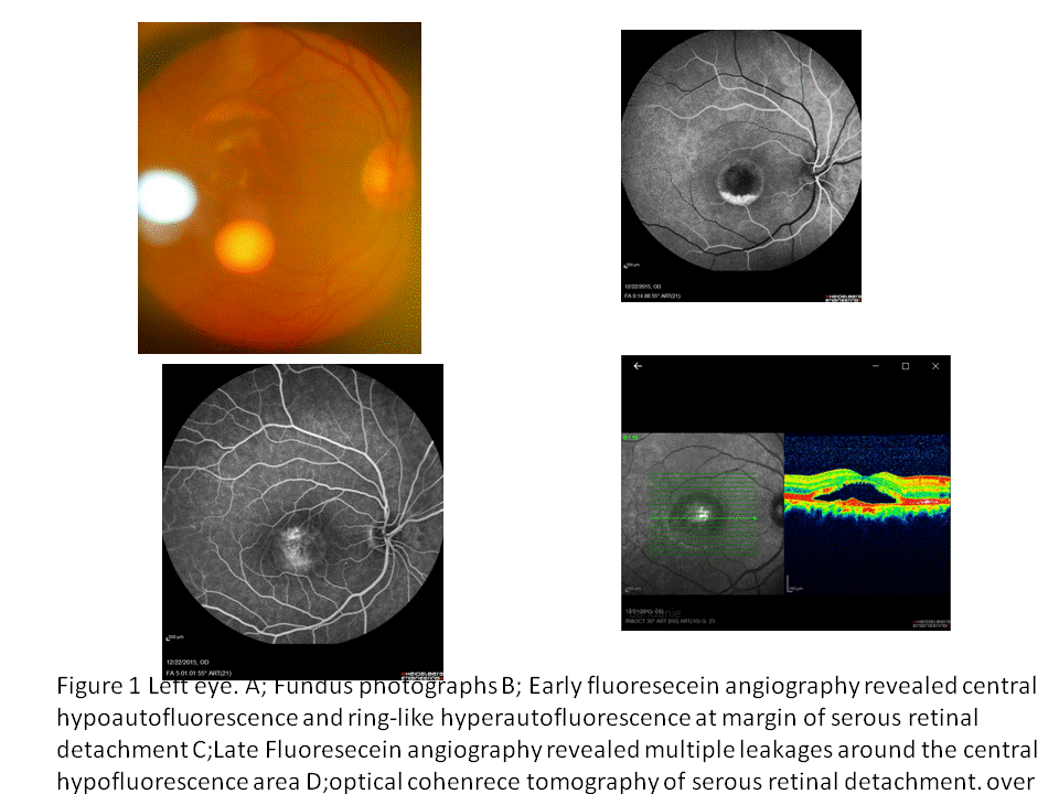
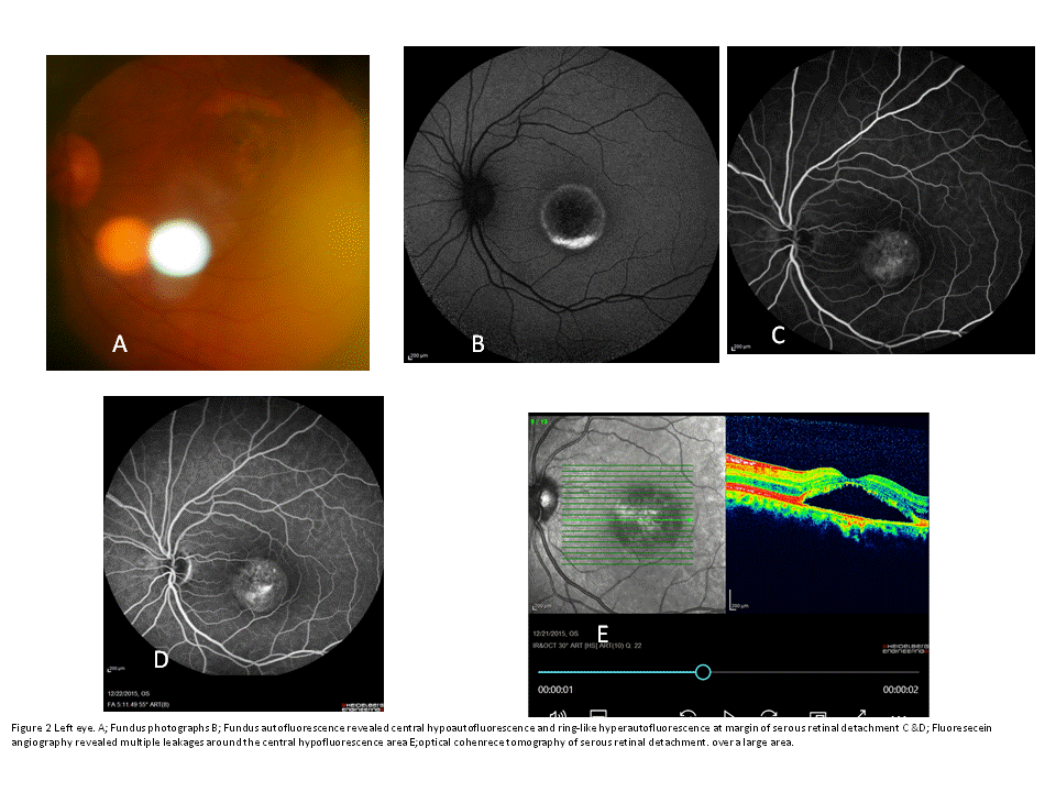
Fifty five years old man came back to eye clinic of driving licenses complain. The patient underwent complete ophthalmic examination. Diagnosis was established according of biomicroscopic examination with macular edema observation, fluorescein angiography with the finding of the point of leakage (Heidelberg digital system) and optical coherence tomography (OCT) demonstrating the detachment of the neuroepithelium of the macula. The best corrected vision in both eye were 20/40. Ophthalmic examination showed serous retinal detachment with well unusual demarcated circum macula lipofushin deposition. The fundous picture indeed of optical coherent tomography and fluorescin angiography showed serous detachment and multiple enhanced fluorescing spot. (Figure 1& 2).
DISCUSSION
This is the characteristic of circumcircular lipofushin accumulation with autoflourescence feature in chronic bilateral central serous chorioretinopathy like feature.
The described multifactorial pathophysiology of CSCR in this paper in spite of advanced retinal imaging but the diagnostic and treatment approach is yet to be defined in future?
In the chronic forms of CSCR, FAF appearance is pathognomonic with multiple oblong descending tracks of decreased FAF, often originating from the optic disc and the macula, that have been referred to as "gravitational tracks "due to their inferior and vertical topography. They are surrounded by a thin contour of increased FAF. The exact significance of these lesions is not elucidated, although it has been suggested that they may result from chronic movements of residual subretinal fluid [3].
Recent imaging findings illustrating subclinical damage put a new rule on the ophthalmic community to develop treatments that are effective at the earliest sign of disease to prevent photoreceptor damage [2]. This various clinically bilateral central serous chorioretinopathy was a presentation that a circumcircular characteristic autofluorescence indicating of feature of CSCR like that due to unique feature is reported in this case. FAF shows different characteristics in acute and chronic CSCR so; in acute phase hypo-autofluorescence is predominant because of the blockage by serous effusion. But increased autofluorescence during of chronic CSCR and cases with persistent serous retinal detachment [2] in spite of clinical feature of acute CSCR which is obvious especially in middle-aged men, but the clinical diagnosis in older patients is more challenging importantly in chronic CSCR that often resembles age-related macular degeneration or as a serous retinal detachments masquerading CSC [3-5]
There is suggestion that hypertension, H. pylori infection, steroid usage, sleeping disturbance, autoimmune disease, psychopharmacologic medication use, and Type-A behavior were possible risk factors relating to the occurrence of CSC [2].
Therefore treatment is according of etiology followed by observation. In chronic cases, photodynamic therapy is the mainstay of treatment. Although there is othere modality of treatment including topical or oral treatment, indeed of subthreshold micropulse laser and conventional thermal laser and half-fluence photodynamic therapy may be even more beneficial in treating early cases of CSCRor mineralocorticoid treatments such as oral eplerenone. In cases of acute CSCR, VEGF inhibitors have shown no benefit; however, in cases of chronic CSCR there is some suggestion that anti-VEGF treatment may be beneficial [6].
Although diagnosis of resembling CSCR in this case was obvious clinically, but OCTA is also a helpful tools in differential diagnosis of it as described by de Carlo TE and colugo's that irregular RPEDs in chronic CSCR eyes may harbor neovascularization more often than previously thought [7] The study by Yong Un heterogeneous patterns of hyper FAF are more frequently observed in cases with chronic CSCR than in those with acute CSCR as our finding [8]. The imaging modalities including high-depth spectral domain OCT and auto fluorescence showed specific features that may help in differentiating atypical CSCR from inflammatory and non-inflammatory conditions [9,10].
Indeed the presence of subretinal/intraretinal hyper-reflective dots in OCT, female sex and aged more than 50 years in acute CSCR might help predict the need for early intervention [11].
Diagnosis of certain cases is straightforward in typical cases but, in atypical presentations, CSCR can be a great mimicker and multimodal imaging is then required for the differential diagnosis [12]. Description a case of clinically similarity of bilateral mimicking chronic CSCR with choroidal neovascularization is not possible without paraclinical assay including flourescein angiography, OCT, or OCTA, electero oculography to definitely diagnosis indeed of clinical presentation. The chronic CSCR can mimicking a variety of dystrophic and acquired retinal diseases. Therefore it should be consider in differential diagnosis of such chronic retinal change. In our cases although the foundus photo by photo slit have not obvious feature and artifact masked it more too. By availability of many paraclinical approach it will be knows more of this enigmatic clinical presentation. Differentiation of resembling chronic macular diseases of our cases unfortunately was not possible due to lost patient follow up. However the old age and large lipofusin precipitation in subretinal space could be presume as late stage of best dystrophy that the lipofusin shows a ring of hyperautofluorescence in FAF, a feature rarely seen in CSCR [13].
However, in certain cases of CSCR, the presence of subretinal fibrin may mimic subretinal lipofuscin deposits Hence the precise underlying diagnosis was not possible. The chronic state of CSCR or late stage of best disease or others similar clinical feature with choroidal neovascularisation is much diverse to can determined it. So based our knowledge it is a unique report of an atypical circumcircular macular autoflourescence pattern that can be chronic CSCR with choroidal neovascularization although definite diagnostic need to exclude other mimicking diseases such as viteliform macular dystrophy by electro oculography and other paraclinical finding also.
REFERENCE
- Singh RP, Sears JE, Bedi R, Schachat AP, Ehlers JP, Kaiser PK. (2015). Oral eplerenone for the management of chronic central serous chorioretinopathy. Int J Ophthalmol. 8(2):310–314.
- Liu B, Deng T, Zhang J. (2016). Risk factors for central serous chorioretinopathy: A Systematic Review and Meta-Analysis Retina. 36(1):9–19
- Daruich A, Matet A, Dirani A, Bousquet E, Zhao M, Farman N, et al. (2015). Central serous chorioretinopathy: Recent findings and new physiopathology hypothesis; Prog Retin Eye Res. 48:82-118.
- Tian-Yu Wang, Zhong-Qi Wan, Qing Peng A. (2019). Patient misdiagnosed with central serous chorioretinopathy: A case report. World J Clin Cases. 7(16):2341-2345.
- Liu P, Fang H, An G, Jin B, Lu C, Li S, et al. (2024). Chronic Central Serous Chorioretinopathy in Elderly Subjects: Structure and Blood Flow Characteristics of Retina and Choroid. Ophthalmol Ther. 13:321–335.
- Hunt D, Tyler Bayliss A. (2023). "Workout" for Central Serous Chorioretinopathy. New Retinal Physician. 18-21.
- de Carlo TE, Rosenblatt A, Goldstein M, Baumal CR, Loewenstein A, Duker JS. (2016). Vascularization of Irregular Retinal Pigment Epithelial Detachments in Chronic Central Serous Chorioretinopathy Evaluated With OCT Angiography. Ophthalmic Surg Lasers Imaging Retina. 47(2):128-133.
- Shin YU. (2017). Review and update for central serous chorioretinopathy. Hanyang Med Rev. 37:10-17.
- Kahloun R, Chebbi A, Amor SB, Ksiaa I, Nacef L, Khairallah M. (2014). Multimodal imaging for the diagnosis of an atypical case of central serous chorioretinopathy. Middle East Afr J Ophthalmol. 21(4):354-357.
- Lee YS, Kim ES, Kim M, Kim YG, Kwak HW, Yu SY. (2012). Atypical vitelliform macular dystrophy misdiagnosed as chronic central serous chorioretinopathy: case reports. BMC Ophthalmol. 12:25.
- Lai WY, Tseng CL, Wu TT, Lin HS, Sheu SJ. (2017). Correlation between baseline retinal microstructures in spectral-domain optic coherence tomography and need for early intervention in central serous chorioretinopathy. BMJ Open Ophth. 2:e000054.
- Dubois P, Postelmans L. (2022). Multimodal imaging of atypical central serous chorioretinopathy in a patient with granulomatosis with polyangiitis. J Français d'Ophtalmol. 45(7):e332-e335
- Sahoo NK, Singh SR, Rajendran A, Shukla D, Chhablani J. (2019). Masqueraders of central serous chorioretinopathy. Surv Ophthalmol. 64(1):30-44

