Information Links
Related Conferences
Previous Issues Volume 9, Issue 1 - 2024
Blastocystis hominis as a Causative Agent of Henoch-Schonlein Purpura
Valeri Velev*
Department of Epidemiology and Hygiene, Medical University of Sofia, Bulgaria
*Corresponding author: Valeri Velev, Department of Epidemiology and Hygiene, Faculty of Medicine, Medical University of Sofia, 1431, Zdrave 2 str., Hospital “Mother's home”, Bulgaria, Phone: 0889563412, Email: [email protected]
Received Date: September 02, 2024
Published Date: November 15, 2024
Citation: Velev V. (2024). Blastocystis hominis as a Causative Agent of Henoch-Schonlein Purpura. Mathews J Gastroenterol Hepatol. 9(1):26.
Copyrights: Velev V. © (2024).
ABSTRACT
Blastocystis hominis is one of the most common protozoa in the human intestinal tract. Most authors accept blastocysts as a harmless commensal, but in some cases they are able to cause nonspecific gastrointestinal symptoms. Although very rare, they can be the cause of autoimmune phenomenon.We present a case of a 6-year-old girl with B. hominis in stools, who developed Henoch-Schonlein purpura.
Keywords: Blastocystis hominis, Vasculitis, Diarrhea.
INTRODUCTION
Blastocystis hominis is one of the most commonly described protozoan parasites in human gastrointestinal tract. It is widespread, though the contagion with this microorganism are most often related to stay in tropical or subtropical areas [1]. Most literature consider it a commensal in the colon since it rarely causes symptoms in immunocompetent individuals even with massive infestation. Nowadays, B. hominisis described as a pathogenic protozoa only in immunocompromised patients, in whom, in the event of more severe invasions, it could cause chronic diarrhea syndrome [2,3].
In the colon, B. hominis exists in a variety of forms - vacuolar, multivacuolar, granular, and ameboid form in the event of massive infestation, also occurring in feces, together with the cyst form, which is invasive. It is considered that an avacuolar form does not exist [2].
Henoch-Schonlein purpura (HSP) is a self-limiting, systemic, non-granulomatous, autoimmune inflammatory complex. Its etiology is still not fully elucidated, but infectious agents - bacteria, viruses, parasites, more rarely mediacions, tumour processes, etc., are mainly reported. It is most common in the age range of 4 - 6 years of age - 90% of cases, with slight predominance of male sex [4]. The initiating agent causes formation of antigen-antibody complexes with IgA. These complexes are deposited in the small vessel walls and they activate the complement, which is followed by a cascade of reactions resulting in vasculitis, which can affect many organs and systems - most commonly the skin, the gastrointestinal tract, the kidneys and the joints [5].
CASE REPORT
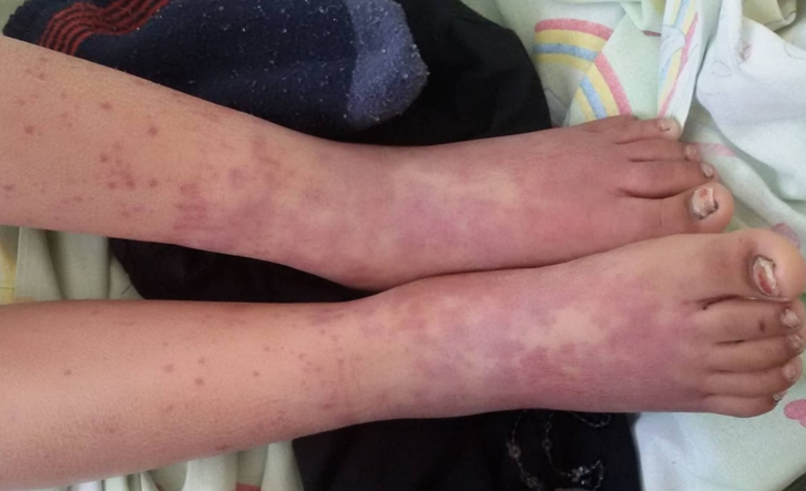
A 6-year-old girl of Roma origin fell ill 3 days before her admission to our clinic with fever, repeated vomiting and watery diarrhea. Prior to hospitalization, she did not take any medications. During the physical examination, the patient was feverish, pale, with dry skin and mucous membranes. The child had complains of diffuse abdominal pain, which was exacerbated upon palpation. Patient’s skin was clean, without rash. Hyperemic throat. Blood tests were performed with the following results: leukocytosis (WBC: 16,900/microL); Hb: 13 g/dL; Hct: 42.1%; BUN: 16 mg/dL; Serum creatinine: 0.8 mg/dL; Urinalysis: no hematuria or proteinuria; ESR: 48 mm/hr; CRP: 6.1 mg/dL; EBV-VCA IgM: Negative; Anti-HAV IgM: Negative; HbsAg: Negative; Anti-HBc IgG: Negative; fecal cultures for pathogen bacteria remained negative. Infusions of glucose and saline solutions and oral rehydration were initiated. Approximately 6 hours after patient’s admission, blood admixtures appeared in her diarrhea stool. Abdominal pain became more severe. At hour 12, palpable hemorrhagic rash appeared on patient’s ankles, and later on her lower legs and gluteal region (Figure 1). Patient’s left ankle became painful and slightly swollen. A fecal test for Campylobacter spp. antigen was carried out, which was negative, and the test for intestinal parasites found cysts and ameboid forms of B. hominis - more than 10 per field. Abdominal ultrasonography found only an insignificant amount of gas in the intestinal loops.
Consultation with a pediatric rheumatologist was carried out, and according to the criteria of the American College of Rheumatology, cutaneous and abdominal form of HSP was diagnosed. Administration of intravenous infusions continued, methylprednisolone 3x20 mg and therapeutic doses of metronidazole were added to the therapy. After three days of treatment, the temperature dropped, the abdominal pain weakened, the rash began to fade. On day 5, the diarrhea syndrome resolved completely, and there were no inflammatory changes in blood counts. No forms of B. hominis were found in the repeated fecal test.
Figure 1. Palpable hemorrhagic rash on the lower limbs of the patient.
DISCUSSION
According to the literature accessible to us, this is the second such case, the first case was reported by Turkish authors in a 30-month-old boy [6]. There is a trend of more and more literature reporting symptomatic infections with B. hominis in immunocompetent individuals [3,7]. The most common presentation is gastrointestinal discomfort, diarrheal diseases, including traveller’s diarrhea [2]. Cases like this, however, demonstrate that this parasite can also cause more serious autoimmune diseases, one of which is Henoch-Schonlein purpura. This once again confirms the need to keep good personal and public hygiene, since infection with B. hominis occurs orally. In all cases with such symptoms - abdominal pain, diarrhea, frequent vomiting - besides microbiological tests, coprological examinations, sometimes three times at intervals of several days, should also be.
CONCLUSION
As time passes and diagnostic capabilities improve, more and more literature sources report symptomatic cases caused by B. hominis. Cases like ours also show possible complications outside the gastrointestinal tract. In view of this, in symptomatic patients with B. hominis isolated from faeces, its role should be discussed.
REFERENCES
- Tan KS, Singh M, Yap EH. (2002). Recent advances in Blastocystis hominis research: hot spots in terra incognita. Int J Parasitol 32(7):789-804.
- Tan KS. (2008). New insights on classification, identification, and clinical relevance of Blastocystis spp. Clin Microbiol Rev. 21(4):639-665.
- Al FD, Hokelek M. (2007). Is Blastocystis hominis an opportunist agent? Turkiye Parazitol Derg. 31(1):28-36.
- Saulsbury FT. (2007). Clinical update: Henoch-Schonlein purpura. Lancet. 369(9566):976-978.
- Roberts PF, Waller TA, Brinker TM, Riffe IZ, Sayre JW, Bratton RL. (2007). Henoch-Schönlein purpura: a review article. South Med J. 100(8):821-824.
- Tutanç M, Silfeler I, Ozgür T, Motor VK, Kurtoğlu AI. (2013). The case of Henoch-Schönlein Purpura associated with Blastocystis hominis. Turkiye Parazitol Derg. 37(2):135-138.
- Chen HH, Liu Q, Deng Y, Zhang HW, Tian LG. (2021). Advances in the research of comorbidity of Blastocystis hominis infections and other diseases. Zhongguo Xue Xi Chong Bing Fang Zhi Za Zhi. 33(5):535-539.
