Previous Issues Volume 9, Issue 5 - 2024
Extensive Cutaneous Larva Migrans: Report of A Case in Yemen
Mohammad Ali Alshami1, Ahlam Mohammad Alshami2, Hadeel Mohammad Alshami1
1Dermatology Department, Faculty of Medicine and Health Sciences, Sana’a University, Sana’a, Yemen
2Department of Conservative Dentistry, Faculty of Dentistry, Sana’a University, Sana’a, Yemen
*Corresponding author: Mohammad Ali Alshami, Dermatology Department, Faculty of Medicine and Health Sciences, Shamlan street 1064/18/10, Sana’a University, Sana’a, Yemen, Telephone: +967-733760082, Fax: +967-1202646, ORCID: 0000-0003-0083-7866, Email: [email protected].
Received Date: June 05, 2024
Published Date: June 21, 2024
Citation: Alshami MA, et al. (2024). Extensive Cutaneous Larva Migrans: Report of A Case in Yemen. Mathews J Case Rep. 9(5):168.
Copyrights: Alshami MA, et al. © (2024).
ABSTRACT
Cutaneous larva migrans (CLM) is the most common helminthic dermatosis, typically found in tropical regions. It is most frequently caused by larvae of the dog or cat hookworm, Ancylostoma braziliense, which can penetrate and migrate through the host's epidermis by releasing degradative enzymes. Diagnosis is generally based on the characteristic clinical presentation, and empirical treatment with oral anthelmintics, such as albendazole and ivermectin, is usually initiated. This report details the case of a 47-year-old man with a 2-month history of an itchy, serpiginous eruption on his back, abdomen, buttock, and thigh. He was successfully treated with oral albendazole 400 mg/day for three days. This is the first documented case of CLM in Yemen.
Keywords: Cutaneous, Larva Migrans, Ancylostoma, Travel medicine, Skin, Zoonotic.
INTRODUCTION
Cutaneous larva migrans (CLM) is most often caused by zoonotic hookworm (Ancylostoma braziliense) larvae, which can penetrate and migrate through the epidermis of the host using degradative enzymes [1,2]. Additionally, CLM is known as creeping eruption, sandworm eruption, plumber’s itch, duck hunter’s itch, and epidermatitis linearis migrans [3]. However, it is most commonly known as creeping eruption because the lesions move as the larvae move in the epidermis. The first report of CLM was described by Lee and published in 1874. Crocker coined the term “cutaneous larva migrans” in 1893. [2] In 1896, Himmelstein proved the parasitic etiology of CLM [4]. In 1929, it was determined that CLM comprises a creeping eruption produced by zoonotic hookworm larvae attributable to Ancylostoma caninum nematode larvae [5]. For many years, the terms “CLM” and “creeping eruption” have been used interchangeably. However, in 2004, Caumes and Danis correctly designated CLM as a syndrome and creeping eruption as a clinical sign [5].
CLM is diagnosed clinically, and treatment comprising oral anthelmintics (albendazole and ivermectin) is administered empirically [6]. Creeping eruption is caused by migrating larvae of zoonotic hookworm and other nematode larvae, adult worms such as Gnathostoma spp. and Loa loa, adult Sarcoptes scabiei (scabies mites), and parasitic fly larvae (migratory myiasis) [7]. Hence, the term “hookworm-related CLM” was introduced to describe CLM caused by the migration of hookworm larvae from dogs and cats within human epidermis [8]. Hookworm-related CLM mainly occurs in resource-poor countries; however, it has been sporadically observed in high-income countries [9]. Tourists who have visited tropical regions have also exhibited hookworm-related CLM. Linear, intensely itchy, and serpiginous tracks that move through the skin are pathognomonic of CLM [9]. These tracks are associated with exposure to hookworm-infested soil or sand. The itchiness is commonly severe and hinders sleep [9]. The larvae remain confined to the epidermis because they cannot penetrate the basal membrane of the human skin; consequently, they cannot complete their lifecycle [9]. Therefore, hookworm-related CLM is a self-limiting disease that is clinically diagnosed. The drug of choice for treating CLM is ivermectin (single dose of 200 μg per kg bodyweight) [9]. However, repeated treatment comprising albendazole (400 mg/day) is a good alternative in countries where ivermectin is unavailable [9]. This report describes the case of a young Yemeni man with extensive CLM and unusual features.
CASE REPORT
A 47-year-old man presented with a 2-month history of intensely itchy serpiginous eruptions on different areas of his body. A cutaneous examination revealed multiple migratory, serpiginous, and curvilinear erythematous lesions on his back, abdomen, buttocks, and thighs (Figure 1a-d). The lesions disappeared within 9 days of initiating oral albendazole treatment (400 mg/day for 3 days). The patient reported walking barefoot in sand contaminated with the feces of dogs and cats. This case was exceptional because the patient presented with multiple tracks, lesions distributed over many body areas, and the presence of simultaneous old and new tracks, which are unusual features that have not been reported previously. In this case report, albendazole was used as the treatment method due to the unavailability of ivermectin. Notably, the efficacy of albendazole in this case appears to be higher than in other reported cases, as the lesions disappeared within just 9 days, whereas it typically takes 4-6 weeks to achieve similar results.
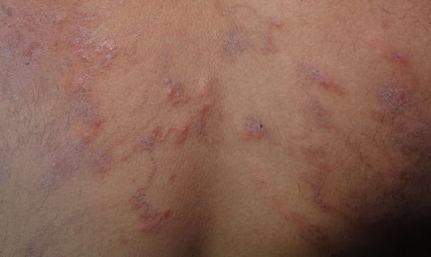
Figure 1a. Upper back close-up, Multiple papulonodules, serpiginous burrows, almost symmetrically bilaterally distributed.
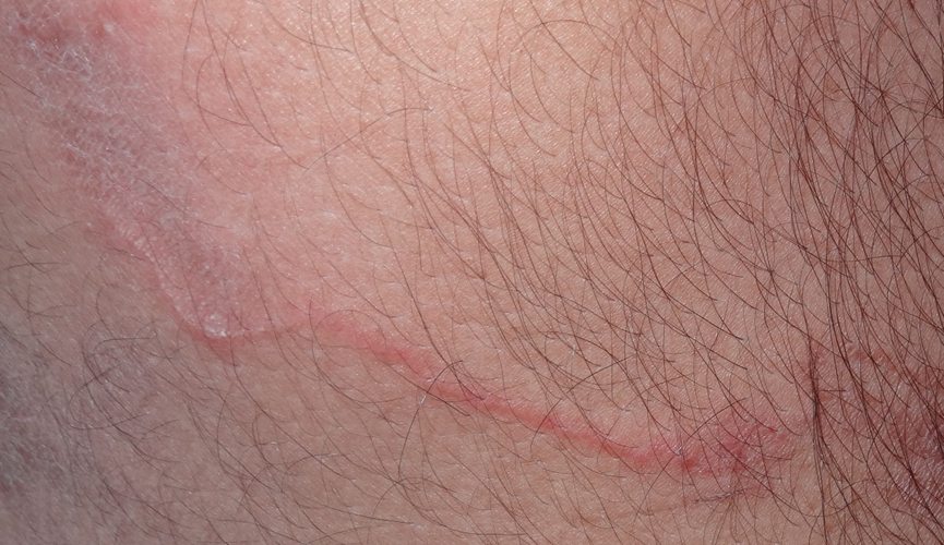
Figure 1b. About 15 cm long erythematous, serpiginous burrow, on the posterolateral aspect of the right side of the back.
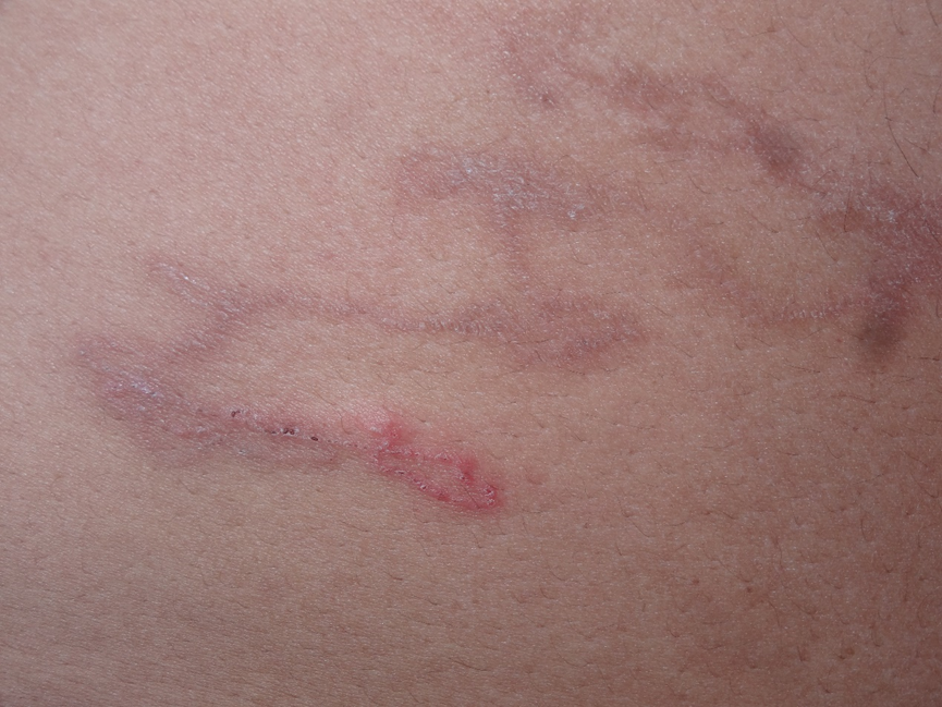
Figure 1c. An about 15 cm long serpiginous burrow is acutely erythematous on its distal end and chronically scaly on the proximal end, seen on the anterolateral aspect of the right side of the abdomen.
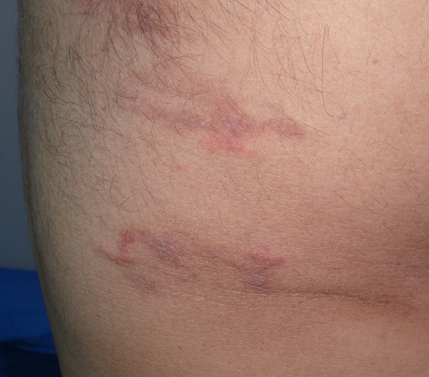
Figure 1d. two serpiginous tracks are visible on the posterolateral aspect of the left thigh.
RESULTS
The lesions disappeared within 9 days of initiating oral albendazole treatment (400 mg/day for 3 days).
DISCUSSION
CLM presents as erythematous, raised, migratory, and serpentine lesions that respond to treatment with anthelmintics [9]. Patients often report a history of walking barefoot in soil or sand infested with hookworm larvae [10]. CLM is commonly observed in tropical and subtropical countries [6]. However, only 20 articles that have included 22 patients with extensive CLM have been published (Table 1). This is the first report of a case of CLM observed in Yemen. CLM is clinically diagnosed when its characteristic serpiginous, migrating tracks are observed. A biopsy is not performed because the parasites are rarely found in the specimens. CLM should be differentiated from diseases caused by other nematode infestations such as larva currens, which is caused by Strongyloides stercoralis and involves rapid track movement (a few centimeters per hour) [11]. Most patients present with one or a few tracks on the extremities or gluteal region after contact with infested soil [7]. Prolonged exposure of large skin surfaces to contaminated soil increases the risk of infection by a large number of larvae, as observed in the present case, resulting in multiple tracks on different sites. Our patient presented with both old and new lesions, suggesting repeated infections and a lack of treatment for more than 2 months. This case demonstrates the need for improved medical knowledge and public education regarding the mode of infestation (skin contact with sand containing larvae of dog or cat hookworms), preventive measures (avoiding walking barefoot in sand contaminated with the feces of dogs and cats), and treatment of CLM.
Table 1. Reported cases of extensive cutaneous larva migrans
|
Year |
1st Author |
Title |
Journal |
Country |
Age &sex |
number |
Sites affected |
|
2001 |
Gourgiotou K |
Treatment of widespread cutaneous larva migrans with thiabendazole |
J Eur Acad Dermatol Venerol |
Greece |
45YF |
1 |
Buttock, foot |
|
2002 |
Karthikeyan K |
Disseminated Cutaneous Larva Migrans |
Indian J Dermatol |
India |
30YM |
1 |
All body except face |
|
2003 |
French SJ |
Severe cutaneous larva migrans in traveler to Jamaica, West Indies |
J Travel Med |
Jamaica |
18YF |
1 |
Chest, breast, back |
|
2004 |
Mehta VR |
Extensive larva migrans |
Indian J Dermatol Venereol Leprol |
India |
26YM |
1 |
Arms, back |
|
2006 |
Malvy D |
Extensive cutaneous larva migrans with folliculitis mimicking multimetameric herpes zoster presentation in an adult traveler returning from Thailand |
J Travel Med |
France |
42YM |
1 |
Abdomen, Flank |
|
2008 |
Jensenius M |
Extensive hookworm-related cutaneous larva migrans in Norwegian travellers to the tropics |
Travel Med Infect Dis |
Norway |
37YM |
1 |
Thighs, hips, genitalia |
|
2008 |
Jensenius M |
Extensive hookworm-related cutaneous larva migrans in Norwegian travellers to the tropics |
Travel Med Infect Dis |
Norway |
29YF |
1 |
Abdomen |
|
2008 |
Tomović M |
Two cases of probable endogenous extensive cutaneous larva migrans in Serbia |
Acta Dermatovenerol Alp Pannonica Adriat |
Serbia |
63YM |
1 |
Abdomen |
|
2008 |
Tomović M |
Two cases of probable endogenous extensive cutaneous larva migrans in Serbia |
Acta Dermatovenerol Alp Pannonica Adriat |
Serbia |
70YM |
1 |
Back |
|
2010 |
Te Booij M |
Löffler syndrome caused by extensive cutaneous larva migrans. A case report and review of the literature |
Dermatol Online J |
The Netherlands |
27YM |
1 |
Both Feet |
|
2011 |
Wambier CG |
Generalized Serpiginous Eruption during Immunosuppressive Treatment for Leprosy Reactive Neuritis |
PLoS Negl Trop Dis |
Brazil |
49YM |
1 |
Abdomen, back |
|
2014 |
Sharma A |
Extensive Cutaneous Larva Migrans. A Case Report |
Indian J Public Health Res Dev |
India |
65YM |
1 |
Abdomen, back |
|
2015 |
González-Ramos L |
Disseminated cutaneous larva migrans infestation |
Semergen |
Spain |
53YF |
1 |
Chest, breasts, arm |
|
2016 |
Comparin C |
Extensive Cutaneous Larva Migrans with Eczematous Reaction on Atypical Localization |
Am J Trop Med Hyg |
Brazil |
32YM |
1 |
Back |
|
2017 |
Rafat M |
Extensive cutaneous larva migrans. A case report |
Journal of Pakistan Association of Dermatologists |
Pakistan |
50YF |
1 |
Right Leg, left thigh |
|
2018 |
Del Giudice P |
Extensive Cutaneous Larva Migrans |
Am J Trop Med Hyg |
France |
18YM |
1 |
Abdomen, chest |
|
2018 |
Tianyi FL |
An unusual case of extensive truncal cutaneous larva migrans in a Cameroonian baby, a case report |
J Med Case Rep |
Cameroon |
9MF |
1 |
Abdomen, chest |
|
2019 |
Gao YL |
Cutaneous larva migrans with Löeffler's syndrome |
Am J Trop Med Hyg |
China |
26YF |
1 |
Right arm, thighs |
|
2019 |
Gunawan H |
Bullous Cutaneous Larva Migrans and Generalized Cutaneous Larva Migrans. A Rare Clinical Manifestation |
Open Dermatol J |
Indonesia |
20YM |
1 |
Scalp, neck, chest, back, buttock, scrotum, extremities |
|
2019 |
Lopes VV |
Disseminated cutaneous larva migrans in a 7-year-old patient |
J. Health Biol Sci |
Brazil |
7YM |
1 |
Right arm, elbow, hand, thigh, knee |
|
2021 |
Ng J |
A Unique Case of Cutaneous Larva Migrans and Asymptomatic Löeffler’s Syndrome |
Cureus |
USA |
52YM |
1 |
Abdomen, foot |
|
2022 |
Tasbun F |
An unusual case of extensive truncal Cutaneous larva migrans from remote area in an Indonesian adult male. A case report |
Indones J Med health |
Indonesia |
21YM |
1 |
Abdomen, back |
CONCLUSION In conclusion, oral albendazole (400 mg/day for 3 days) was effective and safe in treating extensive CLM.
REFERENCES
- Wesołowski R, Mila-Kierzenkowska C, Pawłowska M, Szewczyk-Golec K, Kałużna L, Wozniak AM. (2021). Cutaneous larva migrans imported from a tropical trip - Case report and literature review. Ann Agric Environ Med. 28(4):709-712.
- Sałamatin R, Knysz B, Paszta W, Lelonek E, Matos O, Wesołowska M. (2023). Cutaneous larva migrans: A One Health Perspective on Familial Infection Among Tourists Returning from Southeast Asia. Clin Cosmet Investig Dermatol. 16:3375-3382.
- David A, Srivastava D. (2021). A case report of asymptomatic cutaneous larva migrans. Int J Res Dermatol. 7(5):734-737.
- Sharma R, Singh BB, Gill JP. (2015). Larva migrans in India: veterinary and public health perspectives. J Parasit Dis. 9(4):604-612.
- Caumes E, Danis M. (2004). From creeping eruption to hookworm-related cutaneous larva migrans. Lancet Infect Dis. 4(11):659-660.
- Purdy KS, Langley RG, Webb AN, Walsh N, Haldane D. (2011). Cutaneous larva migrans. Lancet. 377(9781):1948.
- Heukelbach J, Feldmeier H. (2008). Epidemiological and clinical characteristics of hookworm-related cutaneous larva migrans. Lancet Infect Dis. 8(5):302-309.
- Reichert F, Pilger D, Schuster A, Lesshafft H, Guedes de Oliveira S, Ignatius R, et al. (2018). Epidemiology and morbidity of hookworm-related cutaneous larva migrans (HrCLM): Results of a cohort study over a period of six months in a resource-poor community in Manaus, Brazil. PLoS Negl Trop Dis. 12(7):e0006662.
- Feldmeier H, Schuster A. (2012). Mini review: Hookworm-related cutaneous larva migrans. Eur J Clin Microbiol Infect Dis. 31(6):915-918.
- Kaur S, Jindal N, Sahu P, Jairath V, Jain VK. (2015). Creeping Eruption on the Move: A Case Series from Northern India. Indian J Dermatol. 60(4):422.
- Maxfield L, Crane JS. Cutaneous Larva Migrans, in StatPearls. 2024, StatPearls Publishing Copyright © 2024, StatPearls Publishing LLC, Treasure Island (FL).