Previous Issues Volume 3, Issue 1 - 2018
A Case of Post-Lyme Disease Syndrome (PLDS) Involving Motor Neuropathy and Myositis
Yuka Shirai, Yumiko Uchiyama, Masaki Kobayashi, Eiichi Ito, Shinichiro Uchiyama, Yuko Shimizu, Kazuo Kitagawa
Department of Neurology, Tokyo Women's Medical University School of Medicine, Tokyo, Japan
Corresponding Author: Yuka Shirai and Yuko Shimizu, Department of Neurology, Tokyo Women's Medical University 8-1 Kawada-cho, Shinjuku-ku, Tokyo 162-8666, Japan, Tel: +81-3-3353-8111; E-Mail: [email protected], [email protected]
Received Date: 02 Jan 2018 Accepted Date: 17 Jan 2018 Published Date: 19 Jan 2018 Copyright © 2018 Shirai Y and Shimizu Y
Citation: Shirai Y, Shimizu Y, Uchiyama Y, Kobayashi M, et al. (2018). A Case of Post-Lyme Disease Syndrome (PLDS) Involving Motor Neuropathy and Myositis . Mathews J Neurol. 3(1): 009.
ABSTRACT
A 53-year-old man presented with bilateral foot drop. His lower-extremity weakness predominantly affected the distal right limb. He presented hypercreatine kinasemia and high antibody titer for Borrelia species (spp). The nerve conduction study and needle electromyography suggested active neurogenic findings, indicating motor neuropathy. The gastrocnemius muscle biopsy showed scattered fiber necrosis and inflammatory cell infiltration, representing myositis. After administration of minocycline, Borrelia spp antibodies became negative. Symptoms gradually improved with repeated intravenous immunoglobulin administration. This is a very rare case of post-Lyme disease syndrome involving motor neuropathy and myositis, which represents an immune-mediated reaction to Borrelia spp infection
KEYWORDS
Post-Lyme Disease Syndrome; Motor Neuropathy; Myositis; Intravenous Immunoglobulin Administration; Borrelia Infection; Intravenous Immunoglobulin.
INTRODUCTION
Post-Lyme disease syndrome (PLDS) is characterized by chronic physical weariness, muscle pain, and neurological deficits, such as paresthesia and memory disturbance, after treatment with antibiotics [1]. The diagnosis of chronic Lyme disease can predominantly be classified into four categories. PLDS is defined as a group of subjective symptoms such as paresthesia and memory disturbance that persist despite proper treatment of Borrelia burgdorferi infection. However, the pathogenesis of PLDS remains unknown. Some reports have described cases of PLDS associated with myositis and neuropathy [2]. Thus,, patients with other conditions may be included [3]. However, we provide the first report of chronic motor axonal neuropathy and myositis without acute symptoms of Lyme disease in a patient with PLDS.
CASE REPORT
A 53-year-old agricultural teacher who traveled frequently both overseas and domestically presented with bilateral foot drop. He had a history of hepatitis B and dengue fever infections. As his job involves teaching agriculture to the local people, he frequently went on walks in the forest, while wear ing short pants, that is, with large areas of exposed skin. He visited many countries, including Sri Lanka and Pakistan. He had also visited many regions in Japan, particularly in Nagano and Hokkaido prefectures. Thus, his risk of exposure to tick bites was very high. He reported that he often felt feverish, but the fevers were not severe. He did not have other notable symptoms.
A year earlier, he first noticed difficulty in his right foot when going up stairs. After 6 months, muscle weakness had expanded to the left foot and was gradually progressing. When he was admitted to our hospital, no skin rash or arthritis was observed, and neither cognitive decline nor cranial nerve system deficits were detected. Fasciculation was evident in bilateral biceps brachii, triceps brachii, and quadriceps femoris muscles. Muscle weakness and atrophy of the lower extremities was bilateral, but was predominantly distal in the right limbs. The grades of manual muscle testing (MMT) were neck flexion, 5; deltoid, 5/5; biceps, 5/5; triceps, 5/5; pectoralis major, 5/5; wrist flexion, 5/5; wrist extension, 5/5; iliopsoas, 3/3; gluteus medius, 4/4; hamstrings, 4-/5; tibialis anterior, 1/3; peroneus longus, 3/5; and extensor halluces longus, 0/0.
He was able to waddle approximately 200 m with the aid of a cane and had a positive Gowers' sign. Tendon reflexes and sensory, cerebellar, and autonomic systems were all normal. Plantar responses were indifferent bilaterally. High levels of serum creatine kinase (CK; 542 IU/l), aldolase, and nonspecific immunoglobulin (Ig) E were seen, and levels of inflammatory markers were normal. Antinuclear and anti-neuronal antibodies such as anti-Yo, anti-Hu, anti-Ri were not detected, except for anti-ganglioside IgG antibodies to GalNAc-GD1a. The antibody titer of anti-GalNAc-GD1a IgG antibody was 0.222, and its optical density cut-off value was 0.1. First, the titer of IgG antibodies to Borrelia species (spp) antigens was found to be high by enzyme immunoassay (EIA) method and was then confirmed by the western blot (WB) method. The examination of cerebrospinal fluid was normal. In nerve conduction studies (NCS), compound muscle action potential amplitudes were reduced in both peroneal nerves, and mildly reduced in both tibial nerves. Motor conduction velocity of both peroneal nerves was delayed, while the results for the upper extremities and all sensory nerves were normal. The tibial F wave latency was delayed (Table 1).
Table 1: Nerve conduction study.
| Nerve Stimulated(Right side) | Stimulation Site | Recording Site | Amplitude�(NL) | Latency(ms)(NL) | Conduction Velocity(m/s) (NL) | F Wave Latency(ms)(NL) |
|---|---|---|---|---|---|---|
| Median(m) | Wrist | APB | 14.2(=6.0) | 3.0(=4.0) | 26.1(=51.0) | |
| Elbow | APB | 14.1(=6.0) | 7.2 | 56.1(=50) | ||
| Ulnar(m) | Wrist | ADM | 12.98(=8.0) | 2.4(=3.4) | ||
| Below Elbow | APB | 14.1(=6.0) | 7.2 | 56.1(=50) | ||
| Above Elbow | APB | 12.49(=8.0) | 7.8 | 43.5(=36) | ||
| Tibial(m) | Ankle | AH | 18.8(=10.0) | 4.8(=5.0) | 53.3(=51.0) | |
| Poplitea | APB | 12.23(=8.0) | 13.4 | 42.1(=45) | ||
| Peroneal(m) | Ankle | TA | 0.14(=10.0) | 9.0(=5.0) | ||
| Headof fibula | TA | 0.09(=8.0) | 18.6 | 37.7(=45) | ||
| Median(s) | Index Finger | Wrist | 18.7(=10.0) | 2.64(=4.0) | 53(=50) | |
| Ulnar(s) | Small Finger | Wrist | 6.70(=6.0) | 2.68(=3.4) | 52.2(=48) | |
| Sural(s) | Calf | Posterior Ankle | 17.40(=8.0) | 2.50(=3.30) | 44(=40) |
Table 2: Needle electromyography.
| nEMG(Right side) | Fibs | PSW | Recruitment | Normal MUP | Polyphasic | High Amp. | Long durat. | Shordurat. |
|---|---|---|---|---|---|---|---|---|
| TibialisAnterior | Moderate | High amount | Discreteactivity | Low amount | High amount | High amount | Low amount | |
| Gastroc. Medial H | Moderate | High amount | Discreteactivity | Low amount | High amount | High amount | Low amount | |
| VastusMedialis | Moderate | High amount | Discreteactivity | Low amount | High amount | High amount | Low amount | |
| Iliopsoas | Moderate | Moderate | Reducedrecruitment | Moderate | High amount | Low amount | Moderate | Low amount |
| BicepsFemoris,S H | Low amount | Low amount | Discreteactivity | Low amount | High amount | High amount | Low amount | |
| Semitendinosus | Moderate | Moderate | Discreteactivity | Low amount | High amount | High amount | Low amount | |
| Trapezius,Upper | Low amount | Low amount | Normal | High amount | Low amount | Low amount | Low amount | |
| BicepsBrachii | Low amount | Low amount | Normal | High amount | Low amount | Low amount | Low amount |
Table 1, 2: Nerve conduction study and needle electromyography
Compound muscle action potential amplitudes were reduced in both peroneal nerves, and mildly reduced in both tibial nerves. Motor conduction velocity in both peroneal nerves was delayed. Active neurogenic findings were seen at TA, Gastroc. Medial H, Vastus Medialis, Iliopsoas and Hamstrings. These were suggestive of motor neuropathy rather than segmental disturbance. †Amplitude: motor in millivolts; sensory in microvolts. Abbreviations: R, right side; L, left side; m, motor study; s, sensory study; NL, normal; APB, abductor pollicis brevis; ADM, abductor digiti minimi; AH, abductor hallucis; TA, tibialis anterior; nEMG, needle electromyography; Fibs, fibrillation; PSW, positive sharp waves; MUP, motor unit potentials; Amp., amplitude; durat., duration; Gastroc. Medial H, medial head of the gastrocnemius muscle; Biceps Femoris, S H, short head of the biceps femoris muscle.
Note: The F wave latency represents the minimum F wave latency.
Abbreviations: Biceps Femoris, S H: biceps femoris short head muscle, CK: creatine kinase, CMAP: compound muscle action potentials, EHL:extensor hallucis longus, EIA: enzyme immunoassay, Gastroc. Medial H: medial head of the gastrocnemius muscle, GC: gastrocnemius muscle, IVIg: intravenous immunoglobulin, MMT: manual muscle testing, TA: tibialis anterior, WB: Western blot
Needle electromyography (nEMG) of the right lower limb muscles showed high amplitude and long duration of motor unit potentials. We also found reduced recruitment and some fibrillation potentials and positive sharp waves at rest, especially in the gastrocnemius muscle. These were not only seen in the tibial nerve area, L5, or S1 area, but in other areas as well. Additionally, there was a diffuse neurogenic change (Table 1). Magnetic resonance imaging (MRI) revealed diffuse inflammation (Figure. 1).
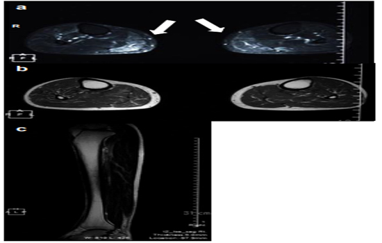
Figure 1: Magnetic resonance imaging (MRI) of the lower limbs, muscle pathology, and clinical course.
a: Short-tau inversion recovery (STIR) MRI of the musculus gastrocnemius (axial, 1.5 T; repetition time, 4050 ms; echo time, 59 ms) shows a diffuse high-intensity area concentrated mainly on the gastrocnemius muscle (arrows). b: Axial T1-weighted image. c: Coronal T2-weighted image.
Irregular size and rounding of the muscle fibers, large group atrophy, scattered fiber necrosis, and inflammatory cell infiltration were detected in the muscle biopsy (Figure. 2).
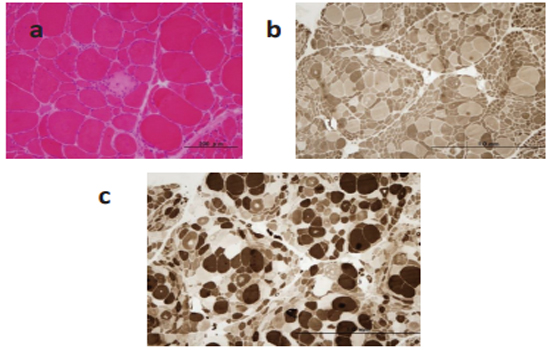
Figure 2: Muscle biopsy from the right gastrocnemius muscle.
Muscle fibers show variation in size and some rounded fibers. Grouped atrophy of type 2 fibers, scattered fiber necrosis, and inflammatory cell infiltration are also present. (a: Hematoxylin and eosin , b: ATPase stain pH9.5, c: ATPase stain pH4.5 )
The MRI of the lumbosacral region showed no abnormalities. Treatment with minocycline was first started, followed by intravenous administration of immunoglobulin (IVIg), and then, oral prednisolone was added. After showing improvement, the patient was discharged. However, his symptoms continued to gradually worsen 1 month after discharge from our hospital. He was only able to walk a distance of 3 m, and thus, was readmitted for further assessments. He was administered a total of four cycles of IVIg repeated every 1-2 months. The EIA for Borrelia Burgdorferi antibodies was negative. As this was the same methodology used at the first examination, administration of minocycline was discontinued. Walking distance gradually improved, and serum CK level normalized (Figure. 3).
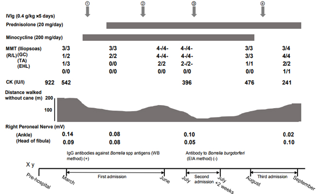
Figure 3: Clinical course.
After admission, the patient was treated with minocycline for 5 months because serum IgG antibody to Borrelia burgdorferi antigens was detected. He was treated with prednisolone (20 mg/day) concurrently. Symptoms improved transiently, and the patient subsequently experienced gradual progression, even after the antibody was no longer detectable. He therefore underwent repeat treatments with intravenous immunoglobulin.
DISCUSSION
This patient demonstrated combined motor axonal neuropathy and myositis without sensory symptoms. There was evidence of myositis and neuropathy by biopsy in addition to high Borrelia spp antibody titers by both EIA and WB methods. Symptoms gradually progressed after the antibody titers to Borrelia spp became negative, and repeated IVIg administrations were required for symptom improvement (Figure. 3). Notably, the motor neuropathy and myositis remained after the antibiotic treatment. PLDS is defined as a syndrome of subjective symptoms persisting despite proper treatment of Borrelia burgdorferi infection; thus, we considered that this was a case of PLDS.
Regarding the differential diagnosis, we considered the following diseases: multifocal motor neuropathy, paraneoplastic syndrome, sarcoidosis, collagen disease, malignancy, and amyotrophic lateral sclerosis. The examination of cerebrospinal fluid was normal, and we did not actively suspect of chronic peripheral dysimmune motor neuropathy, especially multifocal motor neuropathy. Antinuclear and anti-neuronal antibodies, such as anti-Yo, anti-Hu, and anti-Ri, were not detected, and there was no evidence of malignancy. Based on the clinical manifestations, and imaging and laboratory findings, there was no evidence of these diseases.
At the time of the diagnosis, antibodies to Borrelia spp were examined by both EIA and WB methods. However, after antibiotics were administered, we only tested for Borrelia burgdorferi antibodies by the EIA method. In the serodiagnostic standards of the Center for Disease Control and Prevention, Lyme disease is first inspected by EIA or immunofluorescent assay method. If the result is positive, the WB method should be applied. When two of three bands (OspC, BmpA, and Fla) are present for IgM antibody or five of the 10 bands (18kDa, BmpA, etc.) are present for IgG antibody in the WB method, Lyme disease can be diagnosed. The correlation between those epitopes and specific Borrelia spp is not well established. In our case, more than five kinds of positive bands for IgG antibody were detected, and the diagnosis of Lyme disease was established. We could not perform detailed examinations regarding Borrelia spp. However, antibodies to Borrelia burgdorferi proteins were p18, p25, p28, p30, p31, p34, p39, p41, p45, p58, p66, p93, and VlsE by automated immunoblotting. In our case, bands for p41, OspA, b41/I, p41/Igarinii, p41/Iafzeri were strongly positive, and bands for p100, VlsE were slightly positive [4]. Therefore, it was impossible to determine which antigens of Borrelia spp reacted more strongly. Nonetheless, the titer of antibodies to Borrelia burgdorferi proteins was high. Post-treatment examination was negative for the antibodies to Borrelia burgdorferi by EIA, and no further examination was needed. The second EIA evaluation was negative; thus, we concluded that the titers became negative.
The gastrocnemius muscle was especially active in needle electromyography. Therefore, we considered that the MRI findings could reveal further clinical manifestations. Additionally, the tibialis anterior muscle did not present significant changes in the MRI, but we considered that this occurred because the tibialis anterior muscle was affected mainly by neuropathy. In cases of denervation or myositis, the MRI of muscles shows edema-like signal alterations, which indicate a shift of intra- and extra-compartmental fluid. On T2WI, myositis is observed as non-specific findings of increased signals in the involved muscle [5]. Nerve injury induces intrinsic MRI signal alterations on T2-weighted sequences in nerve segments. Thus, we do not consider that these findings are contrary to those expected in myopathic changes [6]. The NCS and nEMG findings indicated motor neuropathy and diffuse neurogenic changes. Although we did not identify a clear myopathic pattern in the nEMG, a few short duration motor unit potentials were detected. As commonly observed in polymyositis, abnormal spontaneous activity, such as insertional activity, fibrillation potentials, and complex repetitive discharges, can be seen in a wide range of myogenic processes. Our patient presented some fibrillation potentials and positive sharp waves, which we considered indicative of myopathic changes. Scattered fiber necrosis and inflammatory cell infiltration shown by gastrocnemius muscle biopsy and hypercreatine kinasemia indicated myositis. Therefore, the muscular weakness in the lower extremities was attributed to the myositis, as well as the motor neuropathy.
Some reports have described myositis or peripheral neuropathy in Lyme disease. Most cases of muscle weakness involved Lyme myositis, with localized pain, swelling, arthritis and normal to moderately elevated serum CK levels, all of which improved with antibiotics. Therefore, these symptoms are considered to be caused by the infection itself [7, 8]. Peripheral neuropathy caused by Lyme disease presents various symptoms such as radiculopathy, mononeuropathy multiplex, and diffuse polyneuropathy [9]. A case of chronic motor axonal polyneuropathy without typical symptoms of Lyme disease and myositis was reported. Symptoms in that case developed even after ceftriaxone treatment and resolved with IVIg [10, 11]. This suggests that an immune-mediated response to Lyme disease caused motor axonal polyneuropathy. In our case, we found a positive low titer of anti-ganglioside IgG antibodies to GalNAc-GD1a. Anti-ganglioside antibodies to GalNAc-GD1a IgG are known to be positive in Guillain-Barre syndrome and other chronic motor neuropathies, but nonspecific positive results with low titer may occur. Because the antibody titer was low in our case, its significance is unknown, but we consider the possibility that some immunological mechanism was at work. This point differs from previous case reports, and it is a feature of our case.
Regarding the treatment, ceftriaxone is generally recommended in late-stage Lyme disease. As this patient wished to take oral medication at that time, we chose minocycline as treatment. Based on the evidence of inflammation in muscle biopsy and the high titers of anti-GalNAc-GD1a IgG antibody, we added prednisolone and IVIg.
To the best of our knowledge, no previous reports of PLDS have been described in conjunction with myositis and motor axonal neuropathy, as seen in our case. Our patient showed antiganglioside antibodies and decreased serum CK levels after IVIg. Therefore, we speculate that not only neuropathy, but also myositis, might be strongly related to the immune-mediated condition in this case of PLDS. Additionally, repeated administration of IVIg may lead to improvement of the symptoms in patients who develop both neuropathy and myositis after antibiotic treatment.
ACKNOWLEDGMENTS
We wish to thank Dr. Hiroki Kawabata (Department of Bacteriology I, National Institute of Infectious Diseases), the Shinjuku Public Health Center, and Dr. Susumu Kusunoki (Department of Neurology, Kinki University) for their valuable information and suggestions. The authors have no competing interests.
CONFLICT OF INTEREST STATEMENT
All authors have no conflicts of interest to declare.
FUNDING ACKNOWLEDGMENT
This work was supported by a Health and Labour Sciences Research Grant on Intractable Diseases (Evidence-based Early Diagnosis and Treatment Strategies for Neuroimmunological Diseases) from the Ministry of Health, Labour and Welfare of Japan.
REFERENCES
- Wormser GP, Dattwyler RJ, Shapiro ED, Halperin JJ, et al. (2006). The clinical assessment, treatment, and prevention of lyme disease, human granulocytic anaplasmosis, and babesiosis: clinical practice guidelines by the Infectious Diseases Society of America. Clin Infect Dis. 43(9): 1089-134.
- Dersch R, Sommer H, Rauer S and Meerpohl JJ. (2016). Prevalence and spectrum of residual symptoms in Lyme neuroborreliosis after pharmacological treatment: a systematic review. J Neurol. 263(1): 17-24.
- Abhishek C, Wormser GP, Marques AR, Latov N, et al. (2011). Anti-Borrelia burgdorferi Antibody Profile in PostLyme Disease Syndrome. Clinical and vaccine immunology. 18(5): 767-771.
- Kumar Y, Wadhwa V, Phillips L, Pezeshk P, et al. (2016). MR imaging of skeletal muscle signal alterations: Systematic approach to evaluation. Eur J Radiol. 85(5): 922-935.
- Stoll G, Wilder-Smith E and Bendszus M. (2013). Imaging of the peripheral nervous system. Handb Clin Neurol. 115: 137-153.
- Holmgren AR and Matteson EL. (2006). Lyme myositis. Arthritis Rheum. 54(8): 2697-700.
- Schoenen J, Sianard-Gainko J, Carpentier M and Reznik M. (1989). Myositis during Borrelia burgdorferi infection (Lyme disease). J Neurol Neurosurg Psychiatry. 52(8): 1002-1005
- Halperin JJ. (2003). Lyme disease and the peripheral nervous system. Muscle Nerve. 28(2): 133-43.
- Rupprecht TA, Elstner M, Weil S and Pfister HW. (2008). Autoimmune-mediated polyneuropathy triggered by borrelial infection? Muscle Nerve. 37(6): 781-785.
- Marques A. (2008). Chronic Lyme disease: a review. Infect Dis Clin North Am. 22(2): 341-60.
- Riccardi VM. (2015). NF1 and the Praxitype. JSM Genetics & Genomics. 2(1): 1006.