Previous Issues Volume 1, Issue 1 - 2017
Aggressive Epidermotropic CD8+ Cutaneous T cell lymphoma: Rare Variant of Cutaneous T cell lymphomas
Dhauna P Karam*, Hosam Hakim, Khalid Al-Khafaji, Pant Mukta Purohit, Bharat Agrawal
Capt James A lovell Federal health care center, 3001 Green bay Road, N.Chicago.
Corresponding Author: Dhauna P Karam, Capt James A lovell Federal health care center, 3001 Green bay Road, N.Chicago.
Tel: 224-610-5554; E-Mail: [email protected]
Received Date: 30 Jan 2017
Accepted Date: 28 Mar 2017
Published Date: 31 Mar 2017
Copyright © 2017 Karam DP
Citation: Karam DP, Hakim H, Al-Khafaji K, Purohit PM, et al. (2017). Aggressive Epidermotropic CD8+ Cutaneous T cell lymphoma: Rare Variant of Cutaneous T cell lymphomas. M J Immu. 1(1): 005.
ABSTRACT
Primary cutaneous aggressive epidermotropic CD8+ cytotoxic T cell lymphomas (AECTCL) are extremely rare of all cutaneous primary T cell lymphomas (PTCL) with less than 1% incidence. A 77 year old male presented to emergency department for evaluation of head to toe rash. He was bit by a wolf spider 5-6 weeks earlier. Patient woke up with spider biting him on his face. Few days later, he developed a maculopapular rash in chest, which failed to respond to OTC ointments. On examination, patient was vitally stable, skin exam revealed multiple confluent erythematous lesions and bullae in the face, neck, back, chest, and extremities. The lesions had central ulceration with necrosis. Punch biopsy of lesion revealed cutaneous aggressive epidermotropic CD8+ cytotoxic T cell lymphoma (AECTCL). Whole body CT/PET scan revealed multiple cutaneous based mass lesions associated with subcutaneous edematous changes. Immunohistochemistry was positive for CD3/CD8/Granzyme B, beta-F1 and TIA-1 further confirming the diagnosis. Patient was treated with gemcitabine for this aggressive variant of T-cell lymphoma. This case highlights a rare and atypical, aggressive variant of cutaneous T cell lymphomas. A constellation of clinical, histopathological, immunophenotypic features aided in our diagnosis.
KEYWORDS
Cutaneous Lymphoma; T Cell; CD8 + Cell; Systemic Chemotherapy; Gemcitabine; Punch Biopsy; Cytotoxicity; and Primary Cutaneous Aggressive Epidermotropic CD8+ Cytotoxic T Cell Lymphomas.
ABBREVIATIONS
PTCL- cutaneous primary T cell lymphomas. AECTCL - Primary cutaneous aggressive epidermotropic CD8+ cytotoxic T cell lymphomas.
INTRODUCTION
Primary Cutaneous T cells Lymphomas (PTCL) are characterized by skin involvement without systemic involvement at the time of diagnosis and disease onset. PTCL accounts for more than 75% of cutaneous lymphomas [1]. 90% of PTCL in western world are classical clinical entities such as mycosis fungoides and its variants, sezary's syndrome on one hand and CD 30 + lymphoproliferative disorders such as anaplastic large cell lymphoma and lymphamatoid papulosis on the other hand. As per the 2016 update of WHO-EORTC classification of tumors of the hematopoietic and lymphatic tissue, there are few other uncommon variants of PTCLs such as subcutaneous panniculitis like T cell lymphoma, extranodal natural killer T cell lymphoma, nasal primary T cell lymphoma, primary cutaneous peripheral TCL, not otherwise classified and rare subtypes. Rare Subtypes of primary cutaneous TCL (PTCL) include: a) Primary cutaneous gamma delta T cell lymphoma, b) Primary cutaneous aggressive epidermotropic CD8+ cytotoxic T cell lymphomas (AECTCL), c) Primary cutaneous acral CD8+ lymphoma and d) Primary cutaneous CD4+ small/medium T cell lymphoproliferative disorder. AECTCL is considered a provisional diagnosis because of its rare incidence and varied presentation [1]. AECTCLs are aggressive variants and are characterized by proliferation of CD 8+ cytotoxic T cells which express CD3/CD8 [2, 3]. In 1991, Berti et al were the first to describe this variant in a 35 year old man [3]. The incidence is 1% of all PTCLs with a male predominance [4]. All proliferating CD 8+ lymphocytes also express a set of toxic proteins such as perforins, granzymes A (GrA), B (GrB), and the T-cell intracellular antigen-1 (TIA-1) that are markers of cytotoxicity [5, 6]. Here we describe a case of primary cutaneous aggressive epidermotropic CD8+ cytotoxic T cell lymphoma (AECTCL), which presented aggressively with a poor outcome.
CASE REPORT
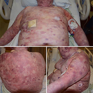
A 77 year old caucasian male presented for evaluation of head to toe rash. He was bit by a wolf spider 5-6 weeks earlier. Few days later, he developed rash in chest, which slowly progressed to become generalized with itching. The patient denied fever or chills. He declined history of recent infections. He thought the rash to be an allergic reaction to spider bite and tried OTC ointments to no avail. Physical examination revealed a vitally stable patient. Multiple confluent erythematous skin lesions and bullae were noted, with central ulceration and necrosis in the face, neck, back, chest, and extremities, sparing palms and soles. There were also many superficial hyperkeratotic plaques and patches [Figure 1, 2, 3].
Basic laboratory investigation including complete blood count and basic metabolic profile were within normal limits. Further workup with HIV/HTLV 1 & 2 testing, hepatitis B and C panel, Epstein-Barr virus (EBV), toxoplasma, rubeola, syphilis, brucellosis, tuberculosis and ricketssiosis panel testing were negative. Autoimmune etiology was ruled out by testing for anti-nuclear antibody, antineutrophil cytoplasmic antibodies (ANCA) and cryoglobulin. Dermatology was consulted and they performed punch biopsy of one of the lesions. Histopathologic examination revealed dense lymphoid cells infiltrating dermis and subcutaneous tissue, which was CD3/CD8 +, Granzyme B + suggestive of AECTCL. Further immunohistochemical testing revealed that the neoplastic cells were positive for CD 7 and BCL- 2, weakly positive for CD 5. The neoplastic cells were negative for CD 20, Pax-5, Bcl- 6, CD 10, CD 30, CD 4, CD 56, CD 34, CD 117, MPO, CD 33 and ALK- 1. Ki-67 was positive in 80% of neoplastic cells. Cyclin- D1 was positive on a small number of lymphocytes but FISH for t (11:14) translocation was negative in peripheral blood ruling out mantle cell lymphoma. Peripheral flow cytometry was negative for sezary's cells ruling out sezary's syndrome. Few other immunohistochemical markers of AECTCL such as TIA-1 and Beta F1 were also positive. TIA -1 revealed dot like staining pattern. Pan CT scan and whole body PET scan was performed with 14.5 mCi of F-18 Fluorodeoxyglucose (FDG). Imaging revealed extensive FDG avid cutaneous lesions. The lesions in external ear and surrounding retro auricular tissues had maximum SUV of 11.7. Soft tissue mass arising from L shoulder and neck were seen, with a SUV of 12.3. Inguinal region lesions had SUV of 15.2. There were some small supraclavicular and axillary lymph nodes with SUV ranging from 6.4 to 10. Patient received gemcitabine chemotherapy for this aggressive variant of T-cell lymphoma.
DISCUSSION
Cutaneous T cell lymphomas are rare malignancies and knowledge about their behavior is limited [7]. Most common of PTCLs are mycosis fungoides and sezary's syndrome (CD 4 + PTCL). The rare PTCL subtypes include four varieties, of which Primary cutaneous aggressive epidermotropic CD8+ cytotoxic T cell lymphomas (AECTCL) are highly lethal malignancies and account for only 1% of all PTCL. Though there are few case reports published on CD 8+ PTCLs [2, 8-10], AECTCL is a clinically unexplored entity. Most CD8+ PTCLs including AECTCL do not respond to treatment and are fatal [2, 4, 9, 10]. Berti et al were the first to describe AECTCL and proposed an earlier classification which evolved over time to the current WHO classification [4, 11-13]. The morphologic features of AECTCL are highly variable. The presentation can involve localized, early patch like lichenoid pattern lesions with marked pagetoid epidermotrophism and subepidermal edema to diffuse dermal infiltrates with nodular tumor-like lesions as seen in our patient. The immunohistochemical findings are positive CD3/CD8, granzyme, perforins, beta- F1and TIA-1. CD 45RA/ CD 45RO, CD2, CD7 may be positive or negative. CD4 and CD5 are usually negative. Genetic testing revealing clonal T cell receptor gene rearrangements are characteristic of CD8+ AECTCL. The diagnosis of AECTCL is based on the constellation of clinical, histopathological and immunophenotypic findings as evident by our case report. Sometimes, the CD4 + lymphomas like Mycosis fungoides and Sezary's syndrome also express CD 8 positivity [14, 15]. Since several PTCLs express CD8+, detailed clinical evaluation with other investigations are needed to diagnose AECTCL. The treatment of AECTCL involves a multimodal approach. Vowels et al in 1994 examined skin biopsies of 12 patients with PTCL and reported presence of Th2 cytokine mRNA in almost all patients [16]. This translates to presence of Th2 helper cell phenotype in PTCL including AECTCLs, which led to development of treatment modalities targeting Th1 response augmentation such as interferons (IFN- alpha, IFN- gamma, IFN-2, IFN-12) and retinoids [17]. Over years, gemcitabine was approved as single agent therapy for patients with untreated AECTCL [18]. Despite its adverse effects, gemcitabine is considered very effective for advanced stages of AECTCL [19]. We used Gemcitabine chemotherapy for our patient and he tolerated it well initially. But over time, he was unable to tolerate therapy and chose hospice care. He died in 9 weeks after diagnosis of AECTCL.
CONCLUSION
Our case highlights the importance of a step wise approach including history and physical exam, followed by investigations in clinical practice that led to diagnosis of a rare clinical presentation. The constellation of clinical, histopathologic and immunohistochemistry help clinch the diagnosis in our patient.
REFERENCES
-
- Willemze R, Jaffe ES, Burg GL. Cerroni, et al. (2005). WHOEORTC classification for cutaneous lymphomas. Blood. 105(10): 3768-3785.
- Agnarsson BA, Vonderheid EC and Kadin EM. (1990). Cutaneous T-cell lymphoma with suppressor/cytotoxic (CD8) phenotype: identification of rapidly progressive and chronic subtypes. J Am Acad Dermatol. 22: 569-577.
- Berti E, Cerri A and Cavicchini S. (1991). Primary cutaneous gamma/delta lymphoma presenting as disseminated pagetoid reticulosis. J Invest Dermatol. 96: 718-723.
- Berti E, Tomasini D, Vermeer MH, Meijer CJ, et al. (1999). Primary cutaneous CD8-positive epidermo-tropic cytotoxic T-cell lymphomas. Am J Pathol. 155: 483-492.
- M Santucci, Pimpinelli N, Massi D, Kadin ME, et al. (2002). Cytotoxic/natural killer cell cutaneous lymphomas: a clinicopathological study of 48 cases from the EORTC cutaneous lymphoma study group. Blood.
- Krenacs L, Wellmann A, Sorbara L, Himmelmann AW, et al. (1997). Cytotoxic cell antigen in anaplastic large cell lymphomas of T- and null-cell type and Hodgkin's disease: evidence for distinct cellular origin. Blood. 89: 980-989.
- E Ralfkiaer, Wollf-Sneedorff A, Thomsen K, Geisler C, et al. (1992). T-cell receptor gamma delta-positive peripheral T-cell lymphomas presenting in the skin: a clinical, histological and immunophenotypic study. Experimental dermatology. 1: 31-6.
- Rodrguez-Pinilla SM, Ortiz-Romero PL, Monsalvez V, Tomas IE, et al. (2013). TCR-? expression in primary cutaneous T-cell lymphomas. Am J Surg Pathol. 37(3): 375- 384.
- Quarterman MJ, Lesher JL, Davis LS, Pantazis CG, et al. (1995). Rapidly progressive CD8-positive cutaneous T-cell lymphoma with tongue involvement. Am J Dermatopathol. 17: 287-291.
- Ohkohchi K, Aiba S and Tagami H. (1986). OKT8-reactive cell mycosis fungoides. Arch Dermatol. 122: 20-22.
- Harris NL, Jaffe ES, Stein H, Banks PM, et al. (1994). A revised European-American classification of lymphoid neoplasms: a proposal from the International Lymphoma Study Group. Blood. 84: 1361-1392.
- Jaffe EF, Krenacs L and Raffeld M. (1997). Classification of T-cell and NK-cell neoplasms based on the REAL classification. Ann Oncol. 8: 17-24.
- Burns MK and Cooper KD. (1993). Cutaneous T-cell lymphoma associated with HIV infection. J Am Acad Dermatol. 29: 394-399.
- Beljaards RC, Meijer CJ, Scheffer E, Toonstra J, et al. (1989). Prognostic significance of CD30 (Ki-1/Ber-H2) expression in primary cutaneous large-cell lymphomas of Tcell origin: a clinicopathologic and immunohistochemical study in 20 patients. Am J Pathol. 135: 1169-1178.
- Kikuchi A, Sakuraoka K, Kurihara S, Akiyama M, et al. (1992). CD8+ cutaneous anaplastic large-cell lymphoma: report of two cases with immunophenotying, T-cellreceptor gene rearrangement and electron microscopic studies. Br J Dermatol. 126: 404-408.
- Vowels BR, Lessin SR, Cassin M, Jaworsky C, et al. (1994). Th2 cytokines mRNA expression in skin in cutaneous Tcell lymphoma. J Invest Dermatol. 103: 669-673.
- Asadullah K, Docke WD, Volk HD and Sterry W. (1998). Cytokines and cutaneous T-cell lymphomas. Exp Dermatol. 7: 314-320.
- Marchi E, Alinari L, Tani M, Stefoni V, et al. (2005). Gemcitabine as frontline treatment for cutaneous T-cell lymphoma. Cancer. 104: 2437-2441.
- Jidar K, Ingen-Housz-Oro, Beylot-Barry M, Paul C, et al. (2009). Gemcitabine treatment in cutaneous T-cell lymphoma: a multicentre study of 23 cases. British Journal of Dermatology. 161: 660-663.